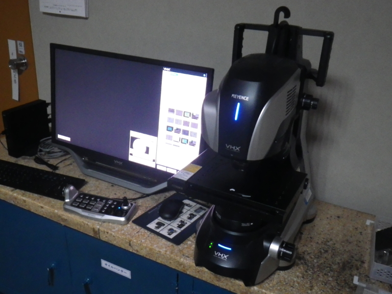
A digital microscope uses an optical lens to magnify an object and a digital camera to capture the image, allowing it to be observed and analyzed on a monitor.
Compared with a stereomicroscope, a digital microscope enables observation at higher magnifications. Adjusting the illumination or combining images can also change how the sample appears.
Captured image data can be used for various types of analysis, including surface roughness and height measurements, as well as 3D image reconstruction.
In this article, we introduce the VHX-X1 Digital Microscope (KEYENCE) used at METEK.
When photographing a glossy product, reflections of the light source, camera, or photographer may appear on its surface. To reduce these reflections, it is often necessary to photograph the product at an angle, making it difficult to obtain a clear image from directly in front of it.
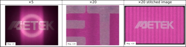
The image-stitching function allows us to capture an image of the entire product without these reflections, provided that the product fits on the stage. The images below are examples we captured.
At 5× magnification, the ring light is reflected on the surface. By stitching together images taken at 20× magnification, we can capture the entire product without this reflection.
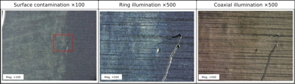
Changing the illumination also changes how the sample appears.
For example, we observed a sample with surface contamination and compared two illumination methods: ring illumination, which produces an appearance similar to observation with the naked eye or a stereomicroscope, and coaxial epi-illumination, which directs light perpendicularly onto the sample along the same optical axis as the camera lens.
The contamination appears whitish under ring illumination and brownish under coaxial epi-illumination. Selecting the appropriate illumination can make features easier to see.
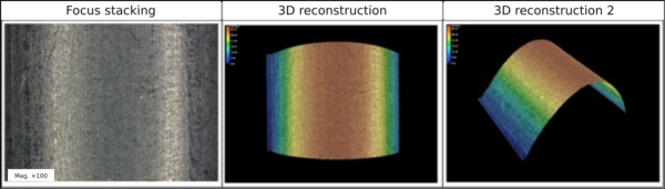
The microscope also has functions for observing three-dimensional objects. Focus stacking produces an image in which the entire surface is in focus, even when the sample has uneven or curved surfaces. The resulting image data can also be used to reconstruct a 3D image.
Measuring the Depth of Surface Features
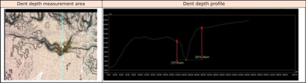
We used the focus-stacking and 3D reconstruction functions to measure the depth of a surface feature.
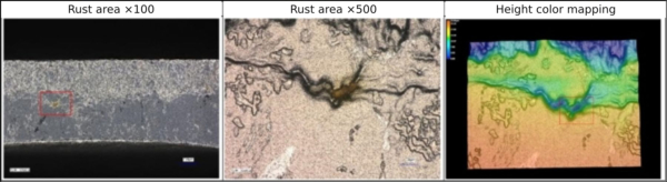
Rust had formed on a plated iron substrate. Observing the rusted area in 3D revealed that it was recessed
The depth of this recessed area was measured at approximately 7 μm.
Observations with the digital microscope suggested the following possible causes of the rust:
1. A deep groove was present, and the plating did not reach the inside of the groove.
2. Moisture entered the groove during the plating process and was not completely removed during drying.
These findings suggest that countermeasures may be needed, such as using a plating process that deposits a coating inside the groove or extending the drying time.
The microscope can measure surface roughness as well as length.
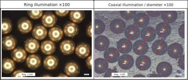
The image shows a diameter measurement of a dummy ball used in plating. Because the outline is difficult to distinguish under ring illumination, we used coaxial epi-illumination for the measurement.
Both profile roughness and areal roughness can be measured. We measured the roughness of a resin sample. Profile roughness is expressed using parameters such as Ra and Rz, while areal roughness is expressed using parameters such as Sa and Sz.
Plating thickness can also be measured by observing a cross section. The image shows a thickness measurement of the copper underplating. The coating can be seen clearly even at high magnification.
The VHX-X1 Digital Microscope enables surface observation with different illumination methods, image stitching, and 3D imaging of three-dimensional objects. It also measures length and surface roughness, making it an essential tool for plating analysis and data collection.
In addition to the functions introduced here, the microscope offers many other capabilities, making it a highly versatile instrument.
At METEK, we use equipment like this in our daily work. If you are considering requesting analysis services, please feel free to contact us.