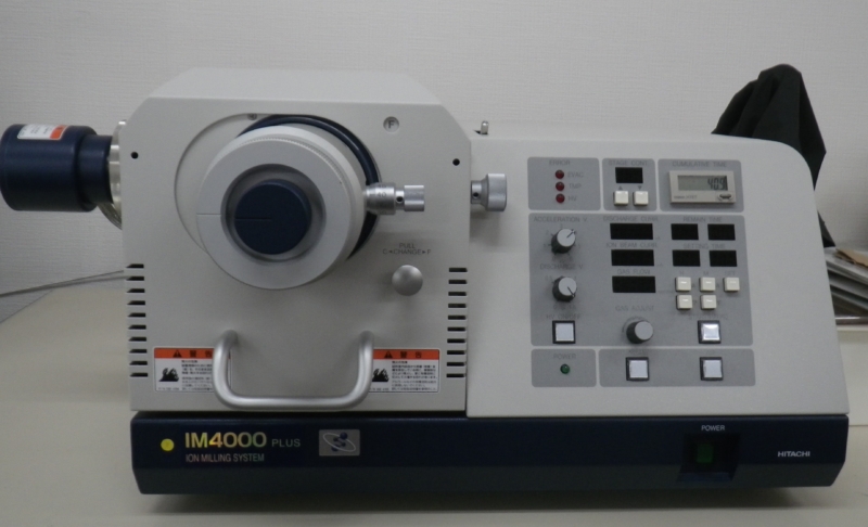
Ion milling equipment is used to prepare sample cross sections for observation with a scanning electron microscope (SEM).
An argon ion beam is directed onto the sample, ejecting atoms from its surface and gradually removing a thin surface layer.
When used after conventional mechanical polishing, ion milling removes edge rounding and fine surface scratches, producing a clearer cross section.
Longer beam exposure allows deeper material removal. Adjusting the beam angle can also accentuate surface relief by taking advantage of differences in etching rates, enabling observation of grain boundaries and multilayer plating structures.
Blistering Analysis Using Ion Milling
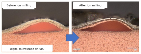
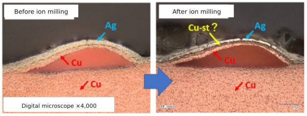
We used ion milling to investigate blistering in a sample with a copper strike and silver plating on an iron-containing copper substrate.
Before ion milling, the blistering could be seen between copper regions. The boundary between copper and silver was visible, but the boundary between the copper substrate and the copper strike was not.
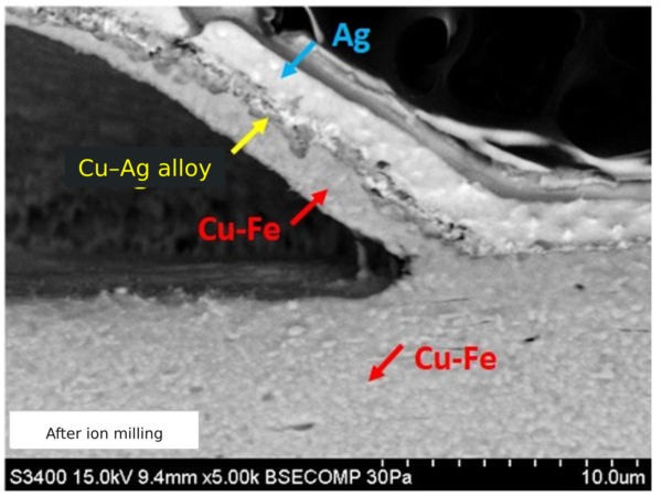
After ion milling, an additional layer appeared to be present between the copper and silver. We initially thought this might be the copper strike layer. The blistering appeared to be occurring within the copper substrate itself.
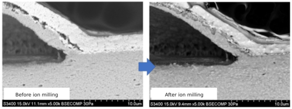
We then examined the sample using SEM for a more detailed analysis.
After ion milling, a layer could be seen between the blistered copper and the silver. However, its boundaries were still not clearly defined.
Energy-dispersive X-ray spectroscopy (EDX) analysis of each layer detected iron in the blistered copper layer, confirming that the blistering had occurred within the copper substrate.
Both copper and silver were detected in the layer between the blistered copper and the silver plating, which we had initially thought was the copper strike. This indicated that it was a copper–silver alloy layer formed during heat exposure.
Because the copper strike was very thin, it appears to have alloyed with the silver, making the boundary indistinct.
In this article, we introduced our newly installed ion milling equipment.
The equipment has made cross-sectional analysis easier, including measurements of plating thickness that cannot be performed using X-ray fluorescence (XRF).
We will continue to use this equipment to improve the quality of our plating and advance our plating technology.